The type of bone defect determines the type of medical care, duration of treatment, and recovery time.
- Fibular Fracture: Symptoms, Treatment and Recovery
- Types of Fibular Fractures
- Types of fibula fractures
- How a doctor treats a fibular fracture
- Treatment of open fibula fractures
- Human Fibula Anatomy Information:
- Which doctors should you see for a fibula exam:
- classification
- Types of lower limb fractures
- Fractures of the articular heads
- Diaphyseal fractures
- Treatment of a fibula fracture
- Rehabilitation after fibular fractures
- First aid
- Treatment methods and remedies
- conservative methods
- surgical treatment
- Medical therapy
- Bones of the lower extremities
- Skeleton of the free part of the lower limbs
- Type B (B1-3)
- Type C (C1-3)
- Symptoms of injury
- Causes of tibia fracture
- Which doctor should I see?
Fibular Fracture: Symptoms, Treatment and Recovery
fibula The fibula and fibula are the long bones of the lower leg. The fibula is small and is located on the outside of the tibia. The tibia is the bone on the inside of the shinbone.
The fibula and tibia are connected to each other at the knee and ankle joints. These two bones help stabilize and support the ankle and foot.
A fracture of the fibula is caused by a fall from a height or a blow to the outer surface of the tibia. A sprained ankle can also lead to a fractured fibula. A fibular fracture can occur anywhere.
Types of Fibular Fractures
Types of fibular fractures include:
- Lateral Fibula Fracture – occurs when the fibula is broken at the ankle joint;
- Proximal fibula head fracture – located at the top of the fibula near the knee joint;
- Avulsion fracture - a fracture in which the tendon tears off part of the bone on the attachment side;
- Stress fractures result from repeated trauma while running or walking;
- Fibula fractures are common in athletes, especially those involved in running, jumping, soccer, and basketball.
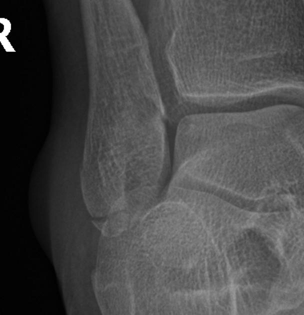
Types of fibula fractures
Fractures of the fibula can occur at the ankle, knee, and mid-shin bone. There are different types of fractures, which can also impact treatment and recovery. The most important types are:
- Lateral fibular fracture – injury to the ankle joint
- Fracture of the head of the fibula, a fracture near the knee
- Avulsion fracture of the fibula, a fracture in which a small portion of the bone is torn out
- Stress fracture of the fibula, a hairline fracture caused by repetitive trauma
- Diaphyseal fracture, a fracture that often affects the middle part of the leg and is caused by a direct impact.
With the exception of stress fractures, the types of fractures mentioned often occur due to trauma or increased pressure on the bone.
How a doctor treats a fibular fracture
Treatment depends on the severity of the fracture, its type, and the location of the injury. Fractures are often classified as:
Regardless of the type of fracture, the traumatologist performs bone fixation and then applies a cast or splint to the leg. This will prevent movement so the fracture can heal.
Closed fibula fractures can, but do not necessarily require, surgery. A splint or cast is usually enough to prevent movement unless other parts of the leg are damaged. If the patient needs additional treatment to realign the bone, the trauma surgeon may prescribe:
- Closed reduction: The doctor aligns the ends of the broken bone without cutting the skin
- Open reduction: The doctor performs an invasive procedure on bones that may be broken in more than two places
- Bone grafting. If the bones do not heal on their own, various procedures and operations are performed, including the insertion of special support pins.
Treatment of open fibula fractures
Rest, ice packs, and elevating the leg are recommended while waiting for treatment. Open fractures require surgery because they can lead to associated injuries such as artery damage.
- Cleaning the wound to prevent contamination and infection
- Stabilizing the wound to keep the bones in place before surgery
- Imaging studies, the results of which make it possible to select a specific type of operation
- Antibiotics to prevent infection.
During surgery, the surgeon may repair the fibula fracture using internal or external fixation techniques. With internal fixation, the doctor inserts metal pins into the broken bone to hold the fracture together while it heals. Severe open fractures require external fixation, in which metal screws or pins protrude above the skin to hold the bone in place.
Human Fibula Anatomy Information:
The fibula (Greek regope), is a thin and long bone with thickened ends. The upper (proximal) epiphysis forms the head, caput fibulae, which articulates with the lateral condyle of the tibia via a flat, round articular surface, facies articularis capitis fibulae. The bony projection, apex capitis fibulae, projects slightly backwards and to the side of this surface.
The body of the fibula is triangular and slightly twisted along its longitudinal axis. The diaphyseal edge of the bone, which faces the tibia and serves to attach the interosseous membrane of the bones, is called the interosseous margin. The lower (distal) epiphysis of the fibula thickens and forms the lateral malleolus (lateral malleolus) with a smooth articular surface (facies articularis malleoli).
Which doctors should you see for a fibula exam:
Are you worried about something? Would you like to learn more about the fibula or do you need an examination? You can make an appointment with dr. – Clinic Eurolaboratory is always there for you! The best doctors will examine you, advise you, provide the necessary care and diagnose the problem. You can also call a doctor at home. clinic Eurolaboratory is open for you around the clock.
How to contact the clinic:
Phone number of our Kiev clinic: (+38 044) 206-20-00 (multichannel). The clinic secretary will find a convenient day and time for you to see a doctor. Click here for our coordinates and directions. Further details on all of the clinic's services can be found on the clinic's homepage.
If you have been examined before Be sure to bring the results with you to the doctor's office. If you have not yet done any examinations, we will carry out the necessary work in our clinic or with our colleagues in other clinics.
It is important that you take a very close look at your general health. There are many diseases that do not initially make themselves felt in our body, but unfortunately are treated too late. To do this, it is simply necessary to be examined several times a year Get a medical check-up several times a yearYour doctor should not only know how to prevent a serious illness, but also how to keep your body and organism as a whole healthy.
If you have questions to ask your doctor, you can find the answers to your questions in our online consultation area. Self Care Tips. If you are interested in reviews of clinics and doctors, you can find information on the forum. You can also register on the physician portal Eurocoolto stay up to date with the latest news and Fibula information on the website, which will then be automatically sent to your mailbox.
classification
Depending on the location, a distinction is made in traumatology and orthopedics:
- Fractures of the tibia in its upper part (fractures of the femoral neck and head of the fibula, fractures of the tibial tuberosity and condyles);
- Fractures of the middle part of the tibia (isolated diaphyseal fractures of the tibia and fibula, diaphyseal fractures of both tibia);
- Fractures of the lower part of the tibia (fractures of the talus bone).
Bone fractures of the upper and lower limbs belong to the group of intra- and periarticular fractures.
Types of lower limb fractures
Fractures of the articular heads
Condylar fractures often occur after a fall from height. They are most commonly split in younger patients and depressed in older patients. A distinction is made between internal and external condylar fractures.
The patient complains of pain and swelling at the injury site. The knee joint is enlarged due to hemarthrosis (collection of blood). A fracture of the external condyle is associated with outward rotation of the tibia, while a fracture of the internal condyle is associated with inward tilt of the tibia. Movement in the joint is extremely painful and restricted. It is difficult or impossible to support the leg. To confirm this, X-rays and MRI of the knee joint are performed.
Treatment:
The shinbone fracture is anesthetized and the joint is punctured if necessary. For condylar fractures without displacement, a plaster cast is applied for 1 month. After immobilization, physiotherapy and exercise therapy are recommended. Full load is allowed after 2 months from the day of injury.
For displaced condylar fractures, reduction is performed and a plaster cast is applied for 6-7 weeks. If the fracture cannot be fused satisfactorily, skeletal traction is performed for up to 2 months. Full weight bearing is possible after 3 months from the day of the injury. Surgical treatment with screws, plates and the Ilizarov device is possible.
Diaphyseal fractures
A diaphyseal fracture of the tibia is the result of direct or indirect trauma. If the interosseous membrane remains intact, there is no displacement of the length of the fragments. An angular shift and a width shift are possible.
The patient experiences pain and swelling in the area of injury. The shinbone is deformed. It is not possible to support the leg. For confirmation, x-rays are taken in two projections.
The fracture site is anesthetized. Displaced fractures are repositioned with a plaster splint for 2 months. Soft tissue interposition (insertion of tissue between fractures) requires surgery.
Treatment of a fibula fracture
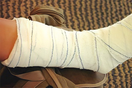
Treatment depends on the type of fracture. If there has been no displacement of the fracture, treatment is not too complicated. The doctor will limit himself to applying a plaster cast starting at the fingertips and ending near the knee joint. Sometimes the cast can be lengthened as needed.
If both tibia bones have been displaced, repositioning is necessary. In the most difficult cases, the doctor can insert spokes into the bone and fix the fracture in the correct position using special metal structures.
The duration of healing of the fibula depends on several factors: the type of injury, qualified and timely first aid, the age of the victim, compliance with the doctor's treatment instructions, etc. In most cases, the healing process takes 2 to 3 months. Callus formation occurs after 6 weeks. In the case of a fracture with displacement and injury to both bones of the knee, the healing process can take longer and take up to 6 months or even longer.
Rehabilitation after fibular fractures
Many patients take the rehabilitation process after the cast is removed lightly and ignore it. This should not be the case because it is an important phase of recovery, without which many complications can arise.
In order for the leg to regain full functionality faster, it is necessary to perform a series of exercises that the doctor recommends. These are usually adapted movements that improve joint mobility and strengthen and strengthen the leg muscles.
A visit to the massage parlor is also not out of the question. If this is not possible, rubbing and kneading should be done at home. Special ointments can be used for this. Wraps, salt baths, waxing and other treatments are also useful. However, it is advisable to consult a doctor before carrying out these treatments to clarify whether they can be used.
First aid
A leg fracture can be simple or clinically complicated and cause significant problems in recovery. It is important to avoid complications from the injury.
In the event of a fracture of the tibia, first aid is provided immediately after the injury to improve the patient's condition during transport to the hospital. Until the paramedics arrive, first aid will be carried out as follows
- Positioning the injured person in the supine position, limiting the mobility of the injured limb;
- Free the injured shin by cutting off clothing and removing footwear as gently as possible;
- cooling the injured limb and administering an anesthetic;
- If the wound area is open, it should be carefully cleaned of dirt and debris;
- if bleeding from an open wound on the lower leg, apply a tourniquet with a time mark;
- Protect the injured leg with hard objects (boards, branches, cardboard).
Correctly performed first aid to the injured person minimizes complications and prevents the patient's condition from worsening.
Treatment methods and remedies
Depending on the type of fracture and its complexity, treatment measures can be carried out using different methods. The choice of techniques depends not only on the type of fracture, but also on the condition and age of the patient.
conservative methods
In practical traumatology, the following procedures are usually used for conservative treatment:
- Fixation method – the repositioning (juxtaposition) of the bone fragments is carried out under local or general anesthesia, then a plaster cast is applied;
- Traction – skeletal traction, which is the slow repositioning of bone fragments using a plaster cast.
These techniques are used when there is no bone displacement or when the fractures can be easily reduced. This procedure is indicated for fractures of the fibular head and neck, as well as transverse fractures of the tibia with small displacement.
surgical treatment
For shin fractures, there are the following indications for prescribing surgery
- Impossibility of closed repositioning of bone fragments;
- inability to hold bone fragments in position;
- Risk of extensive tissue, nerve and vascular damage from bone fragments;
- open fractures;
- Tissue interposition.
The surgical procedure for repositioning bone fragments is carried out using additional methods of immobilization, namely osteosynthesis.
Medical therapy
The therapeutic measures for lower leg fractures always include drug therapy in order to prevent complications, activate bone regeneration and improve the overall condition of the patient. The groups of drugs used in the treatment of lower leg bone fractures include:
- Chondroitin preparations – contain the main components of cartilage tissue and are prescribed in the phase of bone marrow formation;
- Antibiotics – prevent secondary infections and are prescribed for open wounds or surgical procedures;
- Analgesics – relieve pain and improve the general condition of the patient;
- anti-inflammatory medications – reduce swelling and inflammation;
- Calcium supplements and vitamin-mineral complexes – promote better bone regeneration;
- Immunostimulants – increase the body’s defense potential;
- External treatments prescribed after the cast has been removed.
Bones of the lower extremities
Until the age of 12-16, the pelvis (os coxae) consists of three bones connected by cartilage: the hip bone, the pubic bone and the ischium, which fuse together at this age.
The hip bone (os ilium) consists of two sections. The lower, thickened section, the shaft of the hip bone (corpus ossis ilii), is involved in the formation of the hip socket. The upper, wider part is the wing of the hip bone (ala ossis ilii). It is a wide, curved leaf that becomes narrower in the middle. The wing is thickened at the periphery, fan-shaped and ends in the iliac crest (crista iliaca).
The pubic bone (os pubis) has an enlarged part, the body, and two branches. The body of the pubic bone (corpus ossis pubis) forms the front part of the hip socket. The upper branch of the pubic bone (ramussuperior ossis pubis) with the iliopubic eminence, which lies on the connecting line between the pubic bone and the ilium, extends forward from it.
The ischial bone (os ischii) has a thickening (corpus ossis ischii), which closes off the hip socket from below and merges into the ischial branch (ramus ossis ischu) at the front.
[2], [3], [4], [5], [6], [7]
Skeleton of the free part of the lower limbs
The femur is the longest tubular bone in the human body. It has a shaft and two ends. At the upper (proximal) end is the femoral head (Caput femoris), which connects to the pelvic bone.
The tibia consists of two bones. The tibia is medial and the fibula is lateral. Each bone has a body and two ends. The ends of the bones are thickened and have connecting surfaces to the thigh bone (tibia) above and the foot bones below. The space between the shin bones (spatium interosseum cruris) is located between the bones.
The shinbone (tibia) is the thickest bone of the lower limbs. The proximal end of the bone is thickened and forms the medial and lateral condyles (condylus medialis et condylus lateralis). The upper articular surface (facies articularis superior) is directed upwards and connects with the condyles of the thigh.
The fibula is thin and has a fibula head (caput fibulae) at its upper (proximal) thickened end. On the medial side of the head is the articular surface of the fibular head (facies articularis cdpitas fibulae) for articulation with the tibia.
The foot (pes) is divided into 3 parts: tarsus, metatarsus and toes. The skeleton of these parts consists of the tarsal bones (ossa tarsi), the metatarsal bones (ossa metatarsalia) and the toe bones (ossa digitorum pedis).
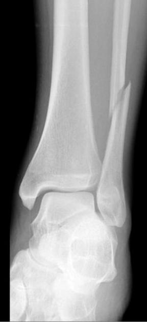
Type B (B1-3)
Ankle: Oblique or torsion fracture that begins at the level of the ankle and runs proximally. The fracture line can be smooth or jagged depending on the force applied.
Medial ankle: Intact or tearing fracture at the base of the ligament, more rarely a tear of the deltoid ligament.
Posterior tibia: Intact or there is a dorsolateral triangular fragment (Volkmann triangle) with tear of the posterior ligament attachments.
Interosseous ligament system: The interosseous membrane is usually intact. The dorsal syndesmosis is more often intact or tears along with the posterior edge of the tibia (Volkmann's triangle).
The anterior syndesmosis (anterior tibiofibular ligament) remains intact below the ankle joint in the event of an oblique fracture of the lateral malleolus. However, if the fracture line begins at the level of the ankle joint gap, the anterior syndesmosis is torn or completely severed. Occasionally, a fracture can tear at the attachment point of the ligament on the tibia (Tillo-Chaputa tuberosity) or fibula. The interosseous membrane usually remains intact.
The severity of the damage to the ligamentous apparatus and the severity of the ankle fracture progressively increases from type A to type B to type C.
In addition to ankle fractures and ligament injuries, fractures of the medial and lateral edges of the talar bone should be found. Both large osteochondral fragments and fractures with cartilage detachment are possible.
Type C (C1-3)
Tibia: Diaphyseal fracture somewhere between the syndesmosis and the head of the fibula.
Lateral malleolus: Tearing fracture or rupture of the deltoid ligament.
Posterior border of the tibia: intact or tear at the syndesmosis insertion site.
Intercondylar ligament apparatus: always torn. Tear in the proximal interosseous membrane of the ankle extending at least to the level of the fibular fracture.
The syndesmosis is torn or detached, along with bone fragments at the attachment site.
Symptoms of injury
The tibia has a tubular structure and is hollow in the middle. The connective tissue cells are arranged in layers and form a compact skeleton. This anatomical structure makes the bone less susceptible to compression and tension, but it is susceptible to twisting and bending. When a force is applied to part of the bone during mechanical trauma, a fracture can occur. Complications such as fractures, dislocations and resulting soft tissue damage are not uncommon. A fracture of the tibia has the following symptoms
- Severe pain at the site of the injury, which may radiate to the thigh and ankle;
- inability to strike foot;
- Restriction of movement of any limb;
- swelling and bruising;
- In case of dislocation, deformation of the leg;
- Characteristic crunching at the time of injury and due to friction of the fragments;
- Abnormal mobility of the bone.
With an open fracture there is also the risk of high blood loss and infection. Connective tissue, bone fragments, damaged ligaments and vessels may be visible in the wound.
Causes of tibia fracture
From an anatomical point of view, the connective tissue of the human legs can withstand very high loads and mechanical stress. But these also have their limits: competitive sports, trauma at work or at home and debilitating illnesses can cause a fracture. The tibia loses its integrity under these conditions:
- Vertical impact;
- excessive pressure;
- twisting of the bone;
- Fracture due to dislocation;
- contact with a hard rib;
- Kick the knee or foot.
A fractured tibia is a particularly dangerous injury for older people. Age-related changes in the body impair coordination of movements, reduce the biochemical composition of connective tissue and lead to a loss of bone strength. Therefore, even a small impact can cause a fracture that takes much longer to heal than in younger people.
Which doctor should I see?
The faster the injured person receives first aid, the faster the main symptoms of the disease - pain, swelling, inflammation - will disappear. It is particularly important to see a specialist if you have complicated injuries. A displaced tibia fracture is treated by a doctor:
Read more:- tibia and fibula.
- syndesmosis.
- The intercondylar syndesmosis is the.
- The lateral ankle is.
- Pain in short fibula when walking.
- Lower third of the fibula.
- function of the fibula.
- Anatomy of the syndesmosis.