Sometimes an X-ray of the heel and a symptom-free foot is taken. This is necessary in order to compare the bones of the affected and healthy leg when the doctor has doubts about the diagnosis.
.png)
- Sever's disease
- Epidemiology/Etiology
- What is a heel bone x-ray?
- Indications for a heel radiograph
- What does an X-ray examination of the heel bone show?
- IMAGES OF THE HEEL BONE
- HEEL PICTURES
- How is the investigation carried out?
- Prices for X-rays of the heel bone
- causes
- Why SM Clinic?
- indications
- Contraindications and limitations
- Indications for X-ray examination of the heel
- Preparing for surgery
- Contraindications to radiographs of the heel bone
- Causes of heel bone fractures
- CT or X-ray for a heel fracture - which is better?
- Causes of heel pain after fractures
- diagnosis
- Preparation for the X-ray examination
- Features of the direct radiograph
- X-ray under stress
- evaluation of results
- Symptoms of a heel bone fracture
Sever's disease
Sever's disease is an abusive syndrome that occurs primarily in young athletes who compete in jumping and running. Other names are heel bone inflammation or Osgood-Schlatter foot syndrome. The disease was first described by James Warren North in 1912. The problem is related to the constant occurrence of micro-trauma from traction on the Achilles tendon. Traction of the tendon occurs at the secondary ossification center of the heel bone, resulting in damage to its apophysis. The cause of this traction apophysitis is repeated micro-injuries or overuse of the heel by young athletes.
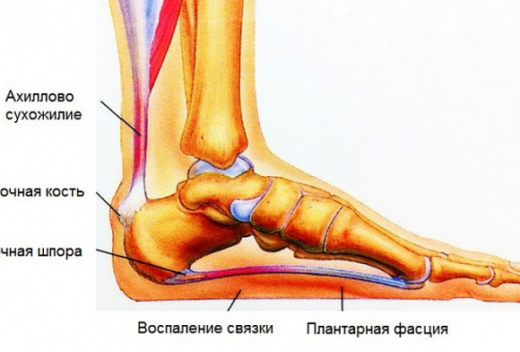
The heel bone is located at the back, plantar part of the foot. The Achilles tendon attaches at the bottom, behind, and about midway through the heel bone. The plantar fascia begins at the medial tuberosity in the plantar area of the heel bone. Closer to the epiphysis is the apophysis, around which the Achilles tendon actually wraps. The growth area of the heel bone and its apophysis are heavily loaded by the plantar fascia and Achilles tendon. In addition, the different growth rates of muscle and bone can lead to a shortening of the tibialis triceps muscle, which can result in less cushioning between the foot and the surface. In addition, the Achilles tendon has a relatively wide insertion area that is anatomically connected to the plantar fascia. This actually helps prevent traumatic rupture of the ossification center.
Epidemiology/Etiology
Sever's disease is an osteochondrosis caused by excessive stress on the heel bone insertion site of the Achilles tendon and the growth plate of the epicondyle. This growth area is C-shaped and can become inflamed if the Achilles tendon is repeatedly pulled. Heel epiphysis inflammation commonly affects young athletes and is believed to be caused by running and jumping.
Friends, Olga Glamazdina's webinar 'Feet: Practice' is coming up. Learn more…
Serious foot disorders are most common in active children and adolescents aged 7-15 years. It occurs particularly frequently during the pubertal growth spurt or at the beginning of the sports season in gymnasts, basketball and soccer players. The disease usually occurs at the beginning of the growing season. Boys are affected more often (ratio 2-3:1).
None of these etiological factors have been prospectively studied. In addition, the available studies have not been conducted systematically, so the reliability or validity of the measurements has not been adequately investigated.
What is a heel bone x-ray?
Of all the bones in the foot, the heel bone is the largest. When walking, running and jumping, it is heavily used, which leads to increased injuries. An X-ray can provide information about the severity of the heel injury, whether there is a fracture, and what type of fracture it is (with or without a dislocation).
Another common problem is heel spurs. It is caused by wearing improper footwear, arthritis or osteoarthritis, but most commonly flat feet. If left untreated, it can lead to complications such as bunions (plantar fasciitis) and fasciitis (inflammation of the foot muscles). The main method of diagnosing these anomalies is X-ray examination, which today can be performed in almost any public or private clinic.
Indications for a heel radiograph
Consider the cases in which a heel x-ray may be recommended:
- Trauma to the foot in the heel area. The causes are different: work trauma, sports injury, fracture as a result of a traffic accident, etc.
- Severe pain in the heel area. Usually occurs in the morning or after a nap when getting up after a short rest and leaning on the heel.
- Pain in the heel area when walking.
- Altered gait (shifting the body's center of gravity to the toes).
- Suspected joint disease – arthritis, osteoarthritis.
- Suspected inflammatory bone disease – osteomyelitis, etc.
- Suspected malignant and benign bone and joint tumors.
What does an X-ray examination of the heel bone show?
X-rays are initially used to assess the integrity of the heel bone. Then its shape and position in relation to the other bones of the foot are analyzed. If heel fractures are detected, their location and direction are examined. If it is a fragment fracture, it is necessary to assess how the fragments are positioned and whether they are displaced.
Tumors on the heel bone can be shown quite well on the x-ray. In this case, the size and location of the tumor must be assessed in order to infer the nature of the abnormality.
IMAGES OF THE HEEL BONE
The aim of the imaging examinations is to examine the shape and structure of the heel bone in various diseases and injuries
Positioning the patient for imaging. X-ray of the heel bone is performed in lateral and axial projection.
A lateral view of the calcaneus is most often obtained from a lateral view of the foot, but sometimes the same patient is placed in the same position to obtain a focused image of the calcaneus by appropriately collimating the X-ray beam to the center of the calcaneus (Figure 451) .
The position for taking an axial radiograph of the heel bone is as follows. The patient lies on his back and stretches out both legs. The foot of the limb in question is in maximum dorsiflexion (Figure 452, a). Sometimes it is withdrawn dorsally with a bandage thrown over the foot, which the patient is holding.
The 13 x 18 cm cassette is laid lengthwise on the table. The foot rests on it with the back of the heel.
The central X-ray beam is cut cranially at an angle of 35-45° to the vertical and directed to the calcaneus tubercle.
The exposure can also be taken with the patient in an upright position. The patient places the soles of the feet of the limb to be removed on the cassette surface, stepping the leg back so that the tibia is at an angle of approximately 45° to the plane of the cassette. The patient should lean against the backrest of a chair in front of them to stabilize their body.
The X-ray beam is aimed at the posterior aspect of the calcaneus tuberosity at an angle of 20° to the vertical (Fig. 452b). .png)
meaningfulness of the images. The lateral projection radiograph shows the structure and contours of the calcaneus and talus (Fig. 453). .png)
The axial view shows the tubercle of the calcaneus and its medial and lateral surfaces (Fig. 454). These images are useful for detecting various pathological changes, fractures, heel spurs (Fig. 455), changes in bone structure, especially after trauma (Fig. 456), etc.
HEEL PICTURES
Determination of the scans. Images of the toes are most commonly used for trauma.
Positioning the patient for X-rays. The toes are clearly visible on straight and oblique x-rays of the foot. If necessary, target images of the toes are taken in the same projections, with the X-ray beam appropriately narrowed and centered on the toe area (Fig. 457, a).
In some cases, x-rays of the fingers are taken from the side by placing a 4 x 5 cm film wrapped in opaque paper under the affected finger. For X-rays of fingers I and V, the foot is placed on the lateral and medial surfaces, respectively, and the X-ray beam is centered perpendicular to the finger in question. When radiographing the fingers I , IIII and IV, the above fingers are pulled down or up with a bandage so that their image does not overlap with the toe joints of the examined finger (Fig. 457, b).
A direct radiograph of the toes clearly shows the phalanges, partially the metatarsals, and the interphalangeal and metatarsophalangeal joints (Fig. 458).
.png)
Tags: foot
234567 Start of activity (date): 30.12.2019 18:33:00
234567 Created by (ID): 989
234567 Keywords: foot, x-ray image, tarsal bone, metatarsal bone, toe bone.
12354567899
How is the investigation carried out?
The examination does not require any special preparation and can be carried out for both acute and elective indications. The area to be examined must be free of any metal objects.
The procedure takes 10 minutes.
X-ray of the heel bone can be done in two ways:
- Straight X-ray - patient is supine with legs bent and soles of feet on table;
- X-ray with weight – the patient stands on the leg being examined and transfers his body weight to the foot.
X-rays of the heel bone are taken in the straight and/or lateral projection.
Prices for the services You can take a look at the price list or call the phone number on the website for details.
The information on this website should not be used for self-medication and self-diagnosis. In the case of acute illnesses, diagnostic tests should only be carried out by a doctor! In order to make a correct diagnosis and prescribe treatment, you should contact your doctor.
Prices for X-rays of the heel bone
- abdomen and pelvis
- X-ray of the abdomen in one projection RUB 3,400.
- X-ray of the kidneys and urinary tract in one projection 4 900 RUB.
- Intravenous (excretory) urography, four exposures, with contrast agent RUB 8,400.
- Retrograde cystography, with contrast agent 9,400 RUB.
- Urethrography, with contrast agent 5,900 rubles
- upper limbs
- X-ray of a finger in two projections 3,400 rubles.
- X-ray of a scapula in one projection 3,900 rubles.
- X-ray of a shoulder blade in two projections 4,400 RUB.
- X-ray of the clavicle in one projection 3,400 rubles.
- Radiography of the clavicle in two projections 4,400 rubles.
- Radiography of the clavicle and shoulder joint in one projection 3,400 rubles
- Radiography of a shoulder joint in a projection 3,400 rubles
- Radiography of a shoulder joint in two projections 4,400 rubles
- Radiography of a shoulder joint in an additional view 2,400 rubles
- X-ray of a shoulder joint in two projections 4,400 rubles
- X-ray of an elbow joint in two projections 4,400 rubles
- Radiography of the bones of the forearm in two projections 4 400 rub.
- Radiography of a wrist in a projection 3,400 rubles.
- Radiograph of a wrist in two projections 4,400 rub.
- X-ray of a hand in two projections 4,400 rub.
- X-ray of two hands in one projection 4,900 rub.
- X-ray of hands with wrists in one projection (determination of bone age) €4,400.
- Head
- X-ray of the skull in two projections 4,400 rubles
- X-ray of the skull in additional and special projections (skull-vertebrae transition, hard palate), one projection 2,400 rubles
- X-ray of the first cervical vertebra (atlas) through the open mouth in a projection 3,400 Rub.
- X-ray of the paranasal sinuses in one projection 3,400 rubles.
- X-ray of the paranasal sinuses in two projections 4,400 Rdnr.
- X-ray of the nasal bone in two projections 3,400 rubles.
- X-ray of the bones of the facial skeleton, orbital cavity in a projection 3,400 Rp.
- X-ray of the Turkish saddle (right and left side projection, 2 images) €4,400.
- X-ray of the cheekbone in two projections €4,400.
- X-ray of the lower jaw in one projection €2,900.
- X-ray of the lower jaw in two projections €3,400.
- thorax
- X-ray of the chest in one projection 2,900 rubles.
- Chest X-ray in two projections 3,400 rubles
- X-ray of the chest in three projections 4,400 rubles
- X-ray of the sternum in one projection 4,400 rubles
- X-ray of the sternoclavicular joints in one projection 3,400 rub.
- X-ray of the ribs (aimed) in one projection 3,400 rubles.
- X-ray of the ribs (aimed) in two projections 4,400 rubles.
- lower limbs
- X-ray of both feet in one projection Rp 4,900.
- X-ray of a foot in two projections 4,900 rub.
- X-ray of the thigh in two projections 4,400 rub.
- X-ray of the tibia in two projections €4,400.
- X-ray of the heel bone in one projection 2,900 rub.
- X-ray of the heel bone in two projections 3,400 rub.
- X-ray of a toe in two projections 2,900 rub.
- pelvic bones and hips
- X-ray of the pelvic bone in one projection €4,400.
- X-ray of a large joint in an additional projection Rp 2,900.
- X-ray of both hip joints in one projection 3,400 rubles.
- X-ray of both hip joints in two projections 4,900 rubles.
- X-ray of both hip joints in three projections €6,400.
- Radiography of a knee joint in a projection 2,900 rubles.
- Radiography of a knee joint in two projections 3,400 rubles
- Radiograph of a kneecap in a 2,900 rub projection.
- Radiograph of a kneecap in two projections 3,400 rub.
- X-ray of an ankle in two projections 3,400 rubles.
- X-ray of both feet under load with radiomorphometry in two projections 5,900 rub.
- X-ray of the sacroiliac joints in two projections 5,400 rub.
- Longitudinal lower limb radiograph, suture 6,900 Rp.
- spine
- X-ray of the first and second cervical vertebrae in one projection €4,400.
- X-ray of the first and second cervical vertebra in two projections €4,900.
- Radiography of the cervical spine in two projections 4,400 rubles
- X-ray of the cervical spine with a functional test 5,400 rubles
- X-ray of the thoracic spine in two projections 4,400 rubles
- X-ray of the sacroiliac joint 4 400 rub.
- X-ray of the lumbosacral spine in one projection 3,400 rub.
- Radiography of the lumbosacral spine in two projections 4,400 rubles.
- X-ray of the lumbar spine with functional tests €5,400.
- X-ray of the sacrum and coccyx in two projections €4,400.
- X-ray of the skull-spine joint in one projection €2,900.
- Digital postural X-ray of the entire spine in one projection €5,400.
- Postural digital X-ray of the entire spine in two projections €6,900.
- X-ray of the lumbar spine and the pelvic bones in children in one projection €4,400.
- mammography
- Mammography of one breast (after mastectomy) 2,900
- Mammography of a gland (focused) in two projections 2,900
- Mammography of both breasts 3,900 rubles
- Ductography of a breast 7,900 rub.
- Radiologically guided interventions
- Capsule Endoscopy
- Capsule endoscopy 80,000 rubles.
- Open
causes
A calcaneus fracture usually results from catatrauma, which is a fall of the body from a height where the feet are resting. In some cases, the bone can also be damaged by a shock wave. In older patients, who show an age-related decrease in bone mineral density, heel fractures can occur even with slight mechanical pressure, such as twisting the foot.
These injuries are called pathologic fractures (resulting from osteoporosis).
Why SM Clinic?
Diagnosis is based on objective examination and an X-ray. From the x-ray, we can clearly see what a traumatic bone injury looks like, where the bone fragments are directed, and what other characteristics the injury has.
In complex clinical cases, CT scans can help make the diagnosis and identify reliable fracture symptoms. With this method, the images are taken layer by layer with minimal increments, allowing the doctor to determine the condition of the tissue in detail.
indications
The main reason for prescribing this test is pain in the foot. The pain occurs most frequently during exertion (e.g. when walking). The patient turns to the doctor who diagnoses plantar fasciitis, a degenerative bone lesion in the heel area caused by stress and overuse. This pathology leads to a compensatory reaction of bone tissue, which leads to the formation of bone outgrowths called osteophytes. This pathology is called heel spurs. It puts increased pressure on the soft tissues of the foot, causing cracks and inflammation in the foot. 'Heel spurs can develop chronic and progressive inflammation of the ligaments and tendons and also damage the muscles around the foot.
Heel spurs mostly occur in middle-aged and elderly people. It also occurs in people who are overweight, have ankle problems, flat feet, or arthritis. He also appears in athletes.
Another common reason for a heel bone x-ray is trauma to the foot. A sharp and violent blow to the foot area can damage the bone - a fracture can occur. Tissue swelling and pain syndromes are noted as a result of torn ligaments and muscles accompanying bone damage.
In the treatment of heel spurs, various physiotherapeutic and instrumental treatments are used to eliminate inflammation of soft tissues:
- Cushions and insoles to relieve pressure on the heel when walking
- Massages, mud and heat treatments
- Ultrasound, laser, shock wave therapy
- Local injection of anti-inflammatory drugs.
X-ray therapy has also proven effective for heel spurs.
To have your heel X-ray in Kiev, but also for other extensive examinations, you can always contact the Meddiagnostika clinic. The clinic has the most modern technical equipment in the form of a digital X-ray machine made in Japan with the possibility of printing the images on tape and storing them on disk for subsequent discussion with a specialist inside and outside the clinic.
Contraindications and limitations
The radiograph of the heel bone is the most important diagnostic tool to establish the diagnosis. It is recommended regardless of the age and gender of the patient.
X-rays of the heels are not recommended for pregnant women without urgent indications (trauma), as it can adversely affect the health of the fetus and the woman herself. In any case, the effects of ionizing radiation are minimized by using special personal protective equipment (aprons, collars, gonad protectors) with which the X-ray laboratory is equipped. When x-raying the heel bone, the x-ray machine has the ability to adjust the radiation field so that the examination area is hit exactly without affecting the rest of the body. The X-ray machine itself also has settings for adults and children.
Indications for X-ray examination of the heel
Due to its special anatomical structure and high daily loads, the heel bone is subject to frequent trauma and pathological processes. An X-ray of the heel is the easiest, fastest and most meaningful way to detect anomalies in time.
An X-ray should be ordered if the following symptoms appear:
- burning or severe pain in the heel
- Restriction of joint mobility in the foot
- pain and discomfort when walking
- Changes in gait, shifting the center of gravity from the heel to the toes
- Swelling, redness and local temperature increase in the area of the heel bone protrusion.
X-rays are taken after trauma to the foot when fractures, breaks, or dislocations are suspected. With bony hypertrophy in the heel area, the diagnostician can assess the development of a spur. X-rays help the doctor assess the effectiveness of treatment for inflammatory diseases, identify the features of bone fusion in fractures, and understand the causes of chronic pain.
Diagnostic X-rays are required if there is a suspicion of:
- osteoporosis
- flat feet
- osteitis
- Achilles tendon rupture and heel bone dislocation
- Arthritis, arthrosis, osteochondropathy.
X-ray shows signs of inflammation characteristic of plantar bursitis, plantar fasciitis, and tendinitis. When describing the x-ray, attention is paid to the integrity of the bony structures, the shape of the heel shaft and its position in relation to the rest of the tarsal area. If abnormalities are found, additional investigations, including examining the other foot, may be needed to make an accurate diagnosis.
Preparing for surgery
X-ray examination of the heel bone for fracture, spur or inflammation is performed without preparation. A referral for the examination may be issued by an orthopedist/traumatologist, podiatrist, rheumatologist, or surgeon. It is advisable to bring the results of previous examinations so that the radiologist can follow the progression of the pathological process.
Immediately before the examination, the specialist will ask you to take off your shoes and free your foot from socks, jewelry and foreign objects.
Contraindications to radiographs of the heel bone
Relative contraindications include pregnancy, lactation and children under 15 years old. Patients in this category are only examined if there is a corresponding indication and after a possible risk has been assessed.
Causes of heel bone fractures
A heel bone fracture is usually caused by a sudden or very strong impact. The severity of the fracture depends on the force of the impact. Some common causes of fractures:
Patients with brittle bones have a higher risk of suffering a heel bone fracture. Bone density may decrease due to the following factors:
- aging
- osteoporosis
- endocrine or intestinal disorders
- corticosteroid treatment
- Lack of regular physical activity
- alcohol and tobacco abuse.
CT or X-ray for a heel fracture - which is better?
CT scans and X-rays are the two main methods used to diagnose injuries to the bony structures of the heel. X-rays provide a very accurate assessment of bone condition and are therefore the preferred form of diagnosis for heel bone fractures. Other benefits of CT and X-rays in fracture diagnosis include:
- The duration of the procedure is only 2-3 minutes;
- an accurate examination without surgical intervention;
- no inconvenience during the examination
- no prerequisites for the patient;
- quick availability of test results.
For these reasons, CT and X-ray are the preferred forms of examination in emergency situations when a surgeon or trauma surgeon needs to act quickly.
Compared to an X-ray, a CT scan of the heel is more detailed and accurate. From a diagnostic point of view, the CT examination has a number of advantages:
- provides a clear and three-dimensional image of the bone;
- three-dimensional bone models can be created;
- the results are immediately visible during the examination;
- The physician can assess both the bony structures and the adjacent soft tissue structures of the limb without the tissue images overlapping.
Causes of heel pain after fractures
For some patients, rehabilitation after an injury takes several weeks. For others, it lasts two months. Doctors recommend not walking again after a broken heel until the pain in the leg has stopped. The symptoms are usually caused by holding the foot in a certain position for a long period of time. This affects the circulation and mobility of lymph and blood and leads to congestion. However, a heel fracture can also lead to complications. This includes:
- post-traumatic flat foot;
- The formation of a false joint;
- heel spur;
- Osteoporosis;
- deforming arthritis of the foot;
- osteomyelitis.
Many patients only experience severe pain after exercising. Others are constantly plagued by it, and the pain intensifies when they rest. Only a doctor can determine what is causing the discomfort and, with an examination, can determine if a heel fracture is present when the limb no longer hurts.
diagnosis
Treatment of a traumatic heel bone injury can be performed using conservative and surgical methods. The choice of one or another tactic depends on the severity of the fracture, the age of the patient and his physiological characteristics. Complications are most common with surgical treatment.
The diagnosis carried out after the heel fracture has been treated, which is essential for determining whether the pain has subsided, is carried out in several steps. It includes an initial examination, a differential diagnosis, and the use of laboratory and instrumental procedures. For example, drugs may be prescribed to patients:
| diagnostic technique | Time |
|---|---|
| X-ray of the ankle | 10 mins |
| Ultrasound of the soft tissues of the foot | 30 minutes |
| electromyography | 300 minutes |
| General blood count and biochemical examination | 10 mins |
MRI is the most powerful examination method to assess the condition of the soft and bony tissues, get a three-dimensional image of the foot and understand when the pain stops after a heel fracture. The cost of the procedure is about 2500-7000 RUB.
Preparation for the X-ray examination
X-ray of the heel bone can be performed without prior preparation. All the patient needs to do is remove shoes and socks from the foot and remove any metal jewelry on the ankle prior to diagnosis. The examination takes place in a separate room in which an X-ray machine is installed.
There are practically no contraindications to an X-ray examination. However, during pregnancy and in small children up to the age of 5-7 years, even low radiation exposure is undesirable. However, if there are no other methods to reliably confirm the diagnosis, then x-rays are not discarded.
Features of the direct radiograph
Routine direct x-rays are taken in the supine position. The examinee is placed on a special table or couch, the legs are bent at the knees, and the foot is positioned so that the entire sole rests on the surface. If the limb is properly positioned and immobilized in this position, the X-ray can be obtained within 1 minute.
X-ray under stress
X-rays of the foot under stress are usually taken to determine the degree of flatfoot. The patient must stand on one limb (the limb to be examined) and place their entire body weight on it. Then the X-ray technician takes the picture.
evaluation of results

X-ray results are interpreted by a radiologist at a private medical center once the diagnosis is made. If a digital machine is used, the X-ray image is sent to a computer, where it can be enlarged to take a closer look at the structures.
In the application, the doctor enters the size of the heel bone, detected lesions, cracks and any fractures. Signs of heel spurs and malignant growths will appear. The diagnosis is made by the attending physician on the basis of x-rays, complaints, visual changes, test data and other examinations.
Symptoms of a heel bone fracture

A calcaneus fracture occurs when a strong external force is applied that exceeds the maximum strength of the bone structure. In older people, injury is possible even with a minor trigger, since osteoporosis develops with age. A broken heel is characterized by severe pain that increases when the foot is touched, swelling of the injured area, and bruising. The calcaneus may appear flattened following the injury and the ability to fully support the foot is severely limited, but mobility in the ankle is preserved.
If a heel bone fracture is suspected, X-rays are taken in three projections. If the diagnosis is confirmed, a plaster cast is applied from the fingertips to the knee joint.
Heel X-ray is a simple diagnostic method used in the treatment of children and adults. The examination in our center can be carried out with modern equipment and without waiting times.
- Anatomy of the foot x-ray.
- Anatomy of the heel bone.
- heel bone injury.
- X-ray of Charcot's foot.
- X-ray anatomy of the ankle.
- Heel bone human anatomy photo and description.
- How Much Does an Orthopedic Surgeon Make?.
- heel bone flexion angle.