4: The nature of the relationship between height and length and foot length. To determine the nature of this relationship, we plotted the foot values on the abscissa and the mean ratios for a given foot length on the ordinate (Fig. 4).
- foot swelling
- Physiological edema
- Advice and guidance from an orthopedist at the Perseus Orthopedic Center. Tel. 8 (495) 469-99-05
- Compensation for the shortened limb
- Results and discussion
- Conclusions
- Causes ↑
- Limb shortening ↑.
- 'Restore the youthfulness of your legs, your legs.'
- Frequently Asked Questions
- useful information
- Anesthesia in bunion surgery
- How to take an X-ray of the foot under proper loading
- How you can benefit from a personal consultation with me
- Different leg lengths Osteopathic treatment
- What is a bent knee?
- Causes of knee deformity
- Transverse flat feet
- causes
- clinical signs
- diagnosis
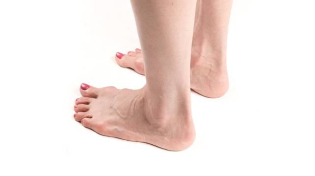
foot swelling
Unfortunately, many people are familiar with foot swelling. Feet swell and become swollen. This happens because excess intercellular fluid accumulates in the tissues. Normally, the amount of fluid in the body is constant: as much as is absorbed, as much should also be released. When fluid is retained in the body (and this excess fluid can be measured in liters), edema occurs. Edema can also be caused by fluid redistribution (abnormal distribution of fluid in the body).
Swelling can occur in various organs and parts of the body, but the feet (especially the ankles and lower legs) are most commonly affected. This is partly because the feet take the most stress (they have to carry all of our weight when we stand or walk, so standing or walking for long periods contributes to foot swelling); second, the feet are the lowest part of our body - gravity helps fluids flow down here and prevents them from draining out.
Swelling on the feet is not always visible. The first symptom may be the appearance of an elastic band around the ankle. In the next stage, the usual shoes become too tight. Women are noticing that the straps of sandals are beginning to 'collapse' and sink into the loose and soft material. Under certain circumstances, the feet can swell even in a healthy person. Swelling usually occurs in the evening as a result of the stress on the feet during the day. However, if the swelling is regular or persistent, you should see a doctor. Edema is a symptom of many different conditions, many of which are very serious and require urgent treatment.
Physiological edema
Swelling in the legs can be physiological, ie it is a consequence of certain circumstances affecting a healthy body and not an expression of an illness. Such causes can be:
- Salty foods. Salt binds water. If you eat a lot of salty foods, especially at night, you may experience puffiness in the morning;
- Alcohol. Alcohol also binds water. That is why a person looks swollen after alcohol abuse. The problems aren't just limited to the face, swollen feet can also occur;
- Hot weather. When it is hot, blood vessels dilate as the body tries to regulate heat exchange. More blood flow means the risk of congestion and swelling in the legs increases;
- prolonged sitting or standing. If the legs remain in one position for a long time, the blood in the legs stagnates, which causes swelling. Sitting cross-legged is the worst. Standing in one place for a long time doesn't help either. Puffiness on the legs is typical for people with standing professions - hairdressers, shop assistants, cooks;
- uncomfortable footwear. When footwear impedes blood circulation in the foot (high heels, tight arches, tight straps), it causes swelling.
Advice and guidance from an orthopedist at the Perseus Orthopedic Center. Tel. 8 (495) 469-99-05
- Orthopedic Surgery Practice
- Advice from a podiatrist
- online consultation
- Appointment with the specialist
- Manufacture of footwear
- orthoses, traction insoles
- Orthoses and braces for the lower limbs
- Stabilization in cerebral palsy
- Insoles for clubfoot.
- With reinforced arch support
- For dynamic splints
- For children
- teenagers
- Adult
- At home
- Footwear for prostheses and aids
- For diabetics
- Tactical shoes, military shoes, outdoor and hiking shoes
- Custom made insoles
1. Orthopedic shoes assembled according to standard lasts
Shoes with insoles that are up to 3 cm shorter
The shoe model is selected by the orthopedist during the visit based on the functional condition and individual anatomical features of the patient's feet.2. Complex orthopedic footwear based on mold and sketch
3. Low shoes with torso compensation (bevel towards the arch of the foot) 4.
Stilettos with adjustable shortening 4.
4a. Insole to compensate for the shortening over the entire range of motion
4b. Insole with partial shortening compensation along the heel + bevel under the heel.5. the shortening compensation along the sole including the patient's shoe.
6. Half-sided insole with shortening compensation (diagonally under the heel) + shortening compensation on the sole 8.
8. Massage, manual therapy
Schedule an appointmentThe design depends on the degree of shortening and is determined by the podiatrist during the consultation
Compensation for the shortened limb
Different limb lengths in the same person have different causes. Shortening of a limb can be congenital, it can be caused by trauma where a broken bone has failed to grow back properly, by bone growth zone disease that causes an occlusion, or by joint disease that results in a flexion contracture. Shortening can affect any of the bones of the lower limbs and adversely affect the overall length of the limb. A shortening of the leg impairs walking. A small difference in length can easily be compensated for by the patient without orthopedic help. Larger limb length discrepancies are usually corrected either through the use of an orthosis or coccyx, or through plantar lengthening of the sole. The orthopedist makes the decision to compensate for the shortening based on the patient's diagnosis.
Hemihypoplasia, congenital hip dislocation, clubfoot, proximal femoral varus, dyschondroplasia, multiple exostoses
Damage to the epiphyseal plate, disruption of the growth zone of the thigh or tibia in osteitis, tuberculosis, arthritis
Osteomyelitis of the thigh or tibia, Brody's abscess, metaphyseal tuberculosis, arthritis, syphilis, elephantiasis, venous thrombosis
Hemangioma, lymphangioma, giant cell tumor, fibrous osteitis, neurofibromatosis, fibrous dysplasia
1. Shortening compensation on the sole of the shoe, also on the patient's shoe

3 Combined shortening compensation
Results and discussion

1. Nature and type of anthropometric relationship between foot length and height. To determine the nature and type of relationship, we plotted the most common foot lengths in newborns and adults on the abscissa axis and the mean height values corresponding to a given foot length on the ordinate axis (Fig. 1). The charts were drawn according to the sex of the subjects.
Figure 1 shows the general trend of foot growth and foot formation: the higher the height, the longer the foot. This pattern is seen in both newborns and adults, regardless of gender. This shows that this anthropometric regularity is inherent in man from birth, regardless of nationality or place of residence, and persists throughout life.
2 The persistence of the anthropometric relationship between foot length and height. Based on the collected data, the degree of correlation between foot length and height was determined. It was 0.73 for men and 0.74 for women and was significantly higher for infants than for adults: 0.81 for boys and 0.84 for girls. These are quite high values, which certainly indicate that there is a connection between body size and foot length.

3 The dimensions of the ratio of height to foot length and their distribution. To determine the degree of proportionality between foot length and height, we plotted the distribution coefficients of their proportions in newborns and adults by sex (Fig. 2).
The diagrams clearly show that the ratio of height to foot length differs between men and women. The mean of this ratio was 6.6 for males and 6.8 for females, with a spread of values greater than 1 % ranging from 6.0 to 7.3 and 6.2 to 7.5, respectively ( within a factor of 1.3).
Conclusions
All the main parameters of the anthropometric relationship between foot length and height, characteristic of adults, are repeated in newborns, but are more pronounced at birth. Differences can only be found in the relative values of the ratio of body size to body length. Such qualitative changes can be explained by the fact that this relationship after birth is influenced by individual personality traits, gender and age differences and other subjective and objective factors that 'blur' and alter the original relationship. The convergence of the fundamental laws of the relationship between height and foot length in newborns and adults indicates that this relationship is already established in the anatomical structure of the human body and is useful in forensic medicine and forensic science for determining height (length) by the foot length can be used.
The authors declare no conflict of interest.
Causes ↑
The main cause is a misalignment or displacement of the pelvis from its natural position, leading to spinal dysfunction. Ultimately, the changes affect the axis of load distribution during movement. Therefore, a displacement, a misalignment of the pelvis is often accompanied by back and neck pain.
Changes in position alter biomechanics, eventually leading to herniated discs, degenerative changes in the vertebrae, osteoarthritis, scoliosis, radiculitis, and narrowing of the spinal canal. In addition, a tilted pelvis gradually causes consequences such as neck pain radiating to the shoulders and arms, and problems with the limbs. Parents may have been diagnosed with hip dysplasia or other joint problems as early as childhood.
osteochondrosis – is a spinal problem in which the vertebral bodies, ligaments, joints and discs of the spine begin to lose their normal function. The main cause of osteochondrosis is a combination of factors affecting the spine over a long period of life. Injuries to the spine and various posture defects also increase the risk of developing osteochondrosis. Some patients attribute the onset of the disease to hypothermia.
Lumbago is a vertebrogenic syndrome. It is manifested by a variety of changes in the lower back. Severe acute pain can occur and the lumbar spine can deform due to muscle tension and tissue pain. Put simply, lumbago is referred to as 'back pain'. If the pain in the lumbar spine is sharp, the affected person can no longer stand up independently and has to adopt a stooped posture.
Limb shortening ↑.
Symptoms of unequal leg length can be mild or severe. The latter contributes to a significant impairment of bodily functions. In the case of moderately severe symptoms, the affected person feels unsafe when walking and frequent falls cannot be ruled out. One hip gets higher than the other.
The most common manifestation is pain:
If If the pelvis is shifted…. If the pelvic obliquity persists over a period of time, the body will compensate for the asymmetry and biomechanical disruption. Over time, the ligaments, tendons, and muscles will adjust. Therefore, treatment can be postponed for a while. Above all, the pelvic obliquity is the most difficult to correct, since a pathological movement pattern develops. The longer the pelvic tilt lasts, the longer it takes for the muscular balance to be restored.
Diagnostics of the lower limbs
It is usually quite easy to determine whether a leg is shortened. While standing, pay attention to the length of the leg. If one leg seems longer than the other, or if the heel keeps stepping on the pant leg while walking, it is most likely short leg syndrome. This pathology contributes to disturbances in the formation of a correct posture, especially in children. If a child has short leg syndrome, they will also develop lower back pain. The pain can spread down the leg to the knee joint and thighs.
Different leg lengths can be well diagnosed with a physical examination. If there is an acute need to diagnose various lesions in the hip joints or spine, instrumental research methods such as MRI or X-rays can be recommended.
Treatment of leg shortening
If you go to an ordinary hospital with these ailments, the prescribed treatment is likely to be ineffective and will not lead to a complete cure, but only to a symptomatic and temporary result. The techniques used in standard orthopedic therapy are not able to relieve the tension in the iliopsoas muscles. The leg will remain short, the joint will remain locked, and the pelvis will remain twisted.
'Restore the youthfulness of your legs
Her legs.'Today there are modern, safe and effective treatments to help you regain the health of your legs. Some of them are completely free, others are very affordable. Unfortunately, many older people do not know about it.
That's why I've created a new, unique campaign for the lovely older generation called 'Restore Youth to Your Feet'.
I invite you to my office for a FREE consultation to examine your feet.
I will also tell you what modern, safe and effective treatments are available to you and how you can get them for free or at the lowest possible cost. We will go through all the options and choose the one that suits you best.
1. Anyone aged 55 and over can participate.
2. You can apply with any foot and ankle problem.
3. The promotion runs from August 1st to September 30th, 2019.
4. You can come to a consultation in person in St. Petersburg at Jaroslawski-Allee 66, Building 1, or get advice online
Call your mothers, grandmothers, relatives, friends - anyone who can benefit from my counseling. Tell as many people as possible, share on your social networks.
Old age is a wonderful time to enjoy life. So don't let pain and discomfort in your feet interfere with your quality of life. Let's work together to make your feet healthy again and make walking your favorite pastime.
If you would like to learn more about the 'Get Your Feet Young Again' campaign, email WhatsApp +79219651182
or call us 8 (812) 336-60-22Frequently Asked Questions
The surgery itself is no different. For all surgeries I perform, I use state-of-the-art medical equipment, the best imported implants, high-quality consumables, the necessary medicines and I use regional anesthesia (a variant of local anesthesia - two injections are given in the foot). There are only two differences in free surgery: 1. If you want to operate on both feet at the same time, you must perform the operation on two different days. The other foot cannot be operated for more than 2 weeks. 2. If you have a quota, you don't have follow-up care I always include follow-up care in the price of the surgeries I pay for. This allows me to monitor how you are feeling, how the bone is healing, how the joint is healing and how your mobility is being restored. This way I can prevent possible postoperative complications. This always leads to excellent results and satisfied patients. Postoperative care can be purchased separately.
useful information
I explain how the treatment works. What to look out for.
How to get the best result.
How you can make your feet healthy and beautiful again and maintain your quality of life for years to come.Anesthesia in bunion surgery
I am often asked two questions: - What anesthesia is used for bunion removal? – Can I choose general anesthesia to be unconscious during the operation?
I answer these two questions in detail in this video, be sure to check it out
How to take an X-ray of the foot under proper loading
Proper assessment of a forefoot deformity (hallux valgus) requires a weight-bearing X-ray, which means the patient must be standing during the exposure.
We recommend the First Diagnostični Center at 10 Sikeirosa Street.
How you can benefit from a personal consultation with me
I am often asked why it is better to come for a consultation before the operation. Finally, you can send photos and social media and go straight to the operating table. I've been operating for 18 years and I've noticed a few things
Different leg lengths Osteopathic treatment
Osteopathy has been listed in the specialist register as the highest level of training for a chiropractic doctor since 2017.
The osteopathic doctor carefully examines the causes of leg length discrepancies when examining a patient. If there is a functional leg length discrepancy, chiropractic treatment is performed.
Osteopathic treatment of leg length discrepancy aims to correct both the immediate cause of limb shortening and the complex changes that have developed in the patient's body as a result. In the case of anatomical shortening, osteopathic treatment is also required, since only an osteopath can restore the correct position of the body structures deregulated by the shortening of the limbs and then adjust the height of the orthosis to the physiology of the body.
It is extremely important to understand that the sooner the disorder is diagnosed and corrected, the less serious compensatory complications appear: scoliosis, pelvic tilt, uneven overload of the ankle, knee, hip and other joints. Therefore, if your child's legs are of different lengths, you should seek qualified help immediately, including the services of a qualified osteopath.
All therapists in our clinic are certified osteopaths with many years of experience. In practice, different leg lengths in both adults and children are corrected during the first therapy session with the osteopath, and then the situation should be monitored over several visits with an interval of 2-3 weeks between therapy sessions. This time is necessary so that a new, stable physiological state of the body can be established.
We are always there for you and your family! SOKLINIKA – Osteopathic family clinic. Clinical institution of the Department of Osteopathy of the Northwest State Medical University named after Mechnikov.
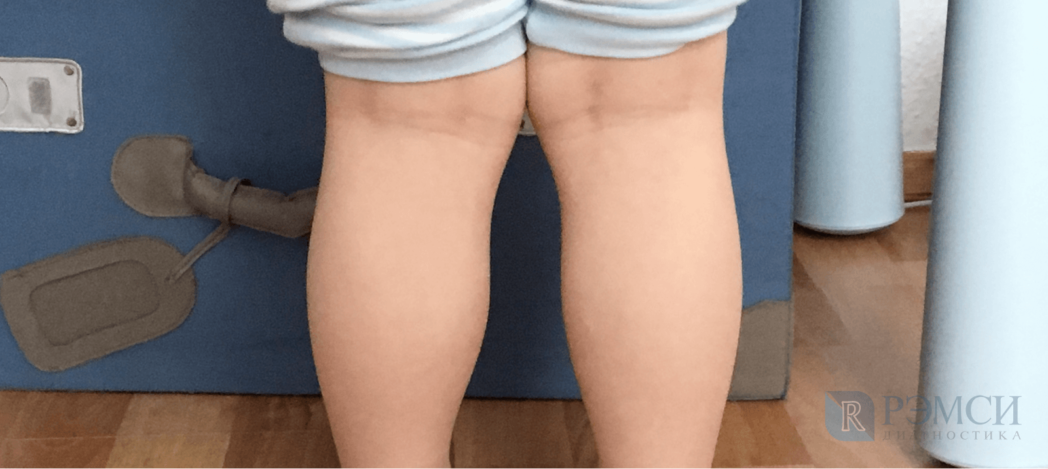
What is a bent knee?
A crooked knee is one of the most common orthopedic disorders. It alters gait and causes clubfoot. There is constant pain in the knees. Even simple walks lead to discomfort and fatigue. A slight deformation does not cause pain in normal life and is purely aesthetic.
With a knee misalignment, the knee moves inward or outward, causing the hip and lower leg to rotate, changing the angle of support, and twisting the leg bones. Doctors distinguish between two types of deformities:
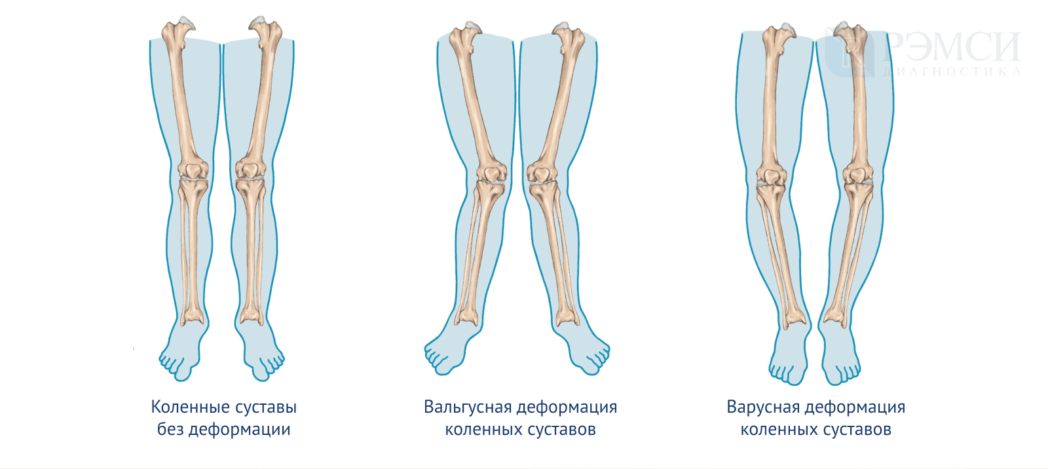
- valgus – where the knees turn inward and the legs look like an X;
- Varus - where the knees move outward and the legs look like an O.
There are also different degrees of severity:
- lightThe deviation of the feet is less than 15 degrees;
- moderateeasy with a leg angle of 15-20 degrees and a shift of the mechanical axis of the leg to the bone edge;
- difficultin which the angle of the lower limbs is more than 20 degrees and the knee deviates from the mechanical axis of the leg.
Causes of knee deformity
The disease can develop at different stages of life: before birth, in childhood and in adulthood. A congenital anomaly of the knee occurs in the mother during pregnancy:
- poisoning by toxic substances,
- frequent stressful situations,
- there was antibiotic therapy,
- there were problems with the endocrine system.
If the baby is born healthy, then there is a chance that joint deformities will develop in the following cases:
- lack of calcium and vitamin D in the diet,
- excessive body weight,
- chronic or congenital cartilage and bone diseases,
- long periods of restricted movement,
- too early attempts to learn to walk.
Bones and cartilage become softer and more brittle due to the lack of nutrients. When a child stands too early on legs that are not yet adjusted to their weight, the joints inevitably become twisted.
In adults, there are several ways to acquire this condition:
- broken legs with dislocations,
- torn ligaments and multiple dislocations of the same knee,
- cartilage damage and disease.
Of course, a lack of vitamins and minerals in the diet can also lead to knee problems in adults. However, when the joints are already well developed, the risk of serious deformation is much lower than in the growth period.
Transverse flat feet
In the name of beauty, women condemn themselves to much suffering. But as we all know, all patience has an end. What's the use of wearing high heels and pumps if the 'bone' protrudes unsightly, widens the foot and literally doesn't allow you to take another step. This foot deformity is caused by a transverse flatfoot.
Our feet are made up of many bones connected by joints, ligaments and tendons. Ideally, the metatarsals (where the pontics attach) should be parallel to each other and held in place by ligaments. In practice, however, these bones are often arranged in a fan shape, which is only acceptable up to a point.
When the supporting bones are too far apart, the following happens: the first metatarsal, to which the big toe attaches, deviates significantly from the rest, and the foot muscles are redistributed. The tendon, previously stretched longitudinally, reverses direction, following the deviated metatarsal and pulling the toe outward. This deviation of the thumb is called hallux valgus. This causes the head of the metatarsal to shift and protrude. Since the shoe is in constant contact with this area, abrasion occurs and the joint becomes inflamed. As a result, osteochondral hypertrophy occurs, popularly referred to as 'bony' hypertrophy.
causes
Transverse flatfoot is most commonly caused by an inherited weakness of the musculoskeletal system. Inheritance occurs in the female line. In men, this form of flatfoot is less common, mainly as a result of trauma. The ratio of affected women to men is 8:2. Standing work (the forefoot is loaded during the day) and uncomfortable footwear exacerbate the problem.
Many people rely on the miraculous effect of supination, which redistributes the load on the foot. However, the disadvantage of supination is that it only relieves the symptoms without eliminating the cause. So how to strengthen the muscles?
In the case of longitudinal flatfoot (when the foot does not have a steep arch and the legs tire quickly), the effect is achieved by strengthening the foot and shin muscles with exercises. In the case of a transverse flat foot, the situation is unfortunately different. The muscles responsible for causing the condition are very small and can be controlled. However, strengthening exercises are useful. By combining foot exercises (left, right, up and down, circular motions) with daily warm baths and foot massage, stubborn bunions can be permanently stopped from growing.
Popular and this recipe: apply a net of 10%iger iodine solution to the 'bone'. This really helps reduce inflammation and thereby stop cartilage growth. Just don't use a more concentrated iodine solution or you could get skin burns. Ditto for vinegar compresses - unfortunately they don't help.
There are many ointments available today that relieve joint inflammation and improve tissue nutrition. However, it is better if the ointment is selected by an orthopedist.
With both forms of flatfoot, it is advisable to wear shoes with a low heel (up to four centimeters). It is advisable to change shoes throughout the day. Wearing shoes at work is uncomfortable and unhealthy. Always remember to wear comfortable, breathable shoes for a change.
When the cartilage that has accumulated at the head of the metatarsal turns into bone, the 'bone' is a constant source of pain. In this case, surgery is the only option.
clinical signs
In pathological conditions, numbness in the legs is a symptom that is always accompanied by a number of other symptoms. Depending on the etiology, these may vary, but the general picture is more or less as follows:
- A feeling of heaviness in the lower limbs;
- Gait disorders;
- inability to distinguish between hot and cold;
- Rapid fatigue, persistent weakness and fatigue;
- tingling and 'goosebumps';
- cramps and increased pain at night;
- Sudden and sudden chest and back pain;
- itching and burning of the skin, sometimes turning blue;
- Severe headache and dizziness.
diagnosis
Thanks to modern instrumental diagnostic methods, the cause can be established relatively quickly. Doctors have the following methods at their disposal:
- X-ray - shows bone deformities, calcifications and other dense formations;
- Computed tomography – allows you to study the bones and substance of the brain and spinal cord and other soft tissues;
- MRI - shows defects in almost all structures; the vascular mode provides an objective view of blood flow;
- Electroneuromyography (ENMG of the lower limbs) – reveals defects in neuromuscular transmission;
- Ultrasound or sonography to show cysts, tumors and other accumulations and to assess the structure of blood vessels;
- General clinical and biochemical blood tests.
The doctors at CELT have a wealth of clinical experience and compare data on various diseases on a daily basis. The specialists are able to find out the exact cause of the ailment and suggest the most effective treatment to improve the situation.
- How to tell if your legs are long.
- A cornea is.
- Shoes that make you taller.
- The child has a shorter leg than the other.
- One leg is shorter than the other.
- Feeling that one leg is longer than the other.
- Leg length proportions.
- difference in leg length.